From Vibration to Sensation: The Neuroscience of Sexual Stimulation
New research on Krause corpuscles explains why a vibrator works where it does, and why an electric toothbrush does not.

After having a phase of “living inside my brain”—getting lost in endless thoughts, memories, “what ifs,” and mental simulations during COVID, stuck in my room for weeks with no physical interaction, I learned how important it is for humans to have environments that provide adequate sensory experiences. After the lockdown, I learned that a good way to stop unregulated thoughts is to consciously focus on external sensory stimuli: the breeze blowing through my hair, the softness of hands, the temperature of water as I soak my feet in a creek. I find senses fascinating—that the same stimulus can be felt differently by different beings, and that every sensory experience starts from receptors at the very end of a nerve.
For the entire semester of the Behavioral and Integrative Neuroscience (BINS for short) class, Professor Anders Nelson jokingly mentioned that the sensory system he was teaching that day was his favorite of the entire course—I found that funny. What is your favorite sensory system, dear readers? Mine is the somatosensory system, which detects and processes bodily sensations including touch, pain, temperature, pressure, and proprioception (position sense). I love it because somatosensory receptors are distributed all over our body, inside and outside. Out of all the different sensations the somatosensory system can detect, one thing that has captured my attention recently is how specific types of touch, particularly vibration, can trigger dramatically different responses depending on exactly where on the body the stimulus lands. Each type of receptor enables us to sense a different parameter of touch, and they are located in precisely the place where they need to be. For instance, you cannot orgasm from the vibration of an electric toothbrush on your tongue, but you can from a vibrator stimulating your genitals. Why? Why does stimulating certain areas of skin in a certain way, for a certain duration, provoke such strikingly different responses compared to other parts of the body? This question was not covered in the lecture, and I suspect it won't be covered in any other lecture either. So I decided to find answers in the newest research on genital sensory mechanoreceptors and share them with readers of the Grey Matters Journal. This article will first go over how somatosensory inputs are processed, then apply that knowledge to the receptors, nerves, and neural circuits related to the senses of the genital area.
ON THE SOMATOSENSORY MECHANORECEPTOR-FOCUSING ON KRAUSE’S CORPUSCLE
Somatosensory end organs are specialized for the functions of the body region or skin type in which they reside. For example, Meissner corpuscles in fingertip skin underlie light touch and support fine motor dexterity, while structures associated with hair follicles respond to hair deflection (Qi et al., 2024). Despite a great deal being known about touch sensors, the physiological properties and functions of sensory structures in the mammalian genitalia had remained completely unclear—until recently.
The genitalia contain specialized sensory structures called Krause corpuscles, first described by anatomist Wilhelm Krause in the 1850s. He noticed they came in two forms: some coiled like a ball of yarn (complex shape), and others small and cylindrical (simple shape). Despite these detailed anatomical descriptions, nobody ever figured out what they actually did or how they functioned. However, a 2024 paper published in Nature by Qi et al. set out to finally answer that question, using mice as a model because they are genetically manipulable and share similar structures with humans.
The first thing Qi et al. (2024) established was where exactly these corpuscles are and how many exist. They took 200-micrometer- thick slices of genital tissue and stained them with two chemical markers: NF200, which lights up large nerve fibers, and S100, which lights up the Schwann cells that wrap around sensory nerve endings to form each corpuscle. This allowed them to see and count every Krause corpuscle precisely under a microscope. What they found was striking. In female genitalia, corpuscles were densely distributed throughout the clitoris but entirely absent from vaginal tissue. In male genitalia, they were found throughout the glans penis and internal prepuce. Despite the size difference between the two organs, the total number of corpuscles in the clitoris and penis was comparable, which means the much smaller clitoris ends up with a 15-fold higher density than the penis. To put that in perspective, the clitoris has three times more Krause corpuscles per unit volume than your fingertips have Meissner corpuscles (Qi et al., 2024). The clitoris is more densely packed with touch sensors than your fingertips. In terms of shape, 93% of clitoral corpuscles were the complex multi-coiled variety, compared to 70% in the penis. In the penis, corpuscles were concentrated in the erectile tissue and the distal tip, and in erect tissue they sat directly adjacent to the blood-filled cavernous spaces, suggesting they may also respond to pressure changes during erection itself (Qi et al., 2024).

ABOUT NEURONAL CONNECTION
Knowing where the corpuscles are is one thing, but to study what they do, the researchers needed to identify which specific neurons supply them, which is not an easy task, since genital tissue contains many different nerve fiber types all mixed together. This is where genetic tools became essential. The researchers used the Cre-lox system, a technique where a gene called Cre is linked to a specific molecular marker, and administering the drug tamoxifen permanently labels only the cells expressing that marker with a fluorescent color, like flipping a light switch exclusively inside those cells. Testing two markers, they found that TrkB+ neurons (labeled by giving tamoxifen to mouse pups five days after birth) terminated in over 90% of all Krause corpuscles in both organs, and nowhere else in genital tissue. Ret+ neurons (labeled during fetal development) innervated approximately 70-80% of corpuscles. complex corpuscles receive input from both TrkB+ and Ret+ neurons simultaneously, while simple corpuscles receive input only from TrkB+ neurons. The team further showed that TrkB signaling is not just a label but a requirement, which means mice engineered to completely lack it had almost no Krause corpuscles in either organ (Qi et al., 2024). No TrkB, no corpuscles.
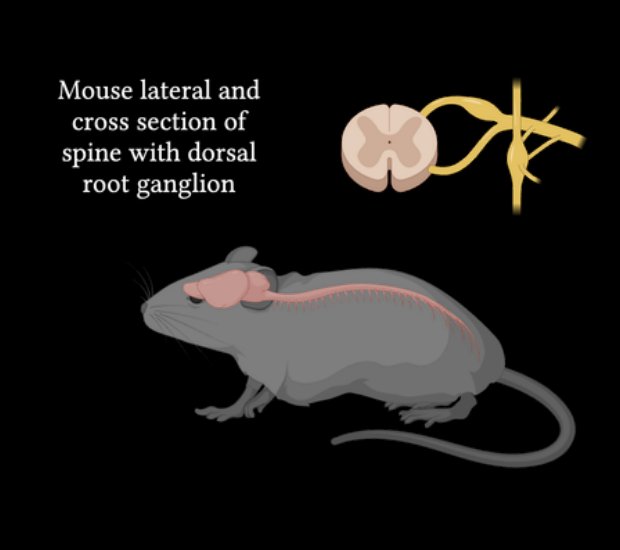
With access to these specific neurons established, the central question could finally be asked: what do Krause corpuscles actually sense? Are they touch sensors, vibration sensors, heat sensors, pain sensors? To find out, the researchers developed an experimental setup to apply precise mechanical and thermal stimuli directly to the external genitalia of living, anesthetized mice while recording individual neuron activity in real time. They surgically exposed the L6 dorsal root ganglion, the cluster of sensory neurons that receives signals from genital tissue and lowered a multielectrode array (MEA) onto it. An MEA is a tiny grid of electrodes that picks up the electrical spikes, called action potentials, that neurons fire when activated. But many different neuron types from many body regions are packed into the same ganglion.
So how do you know which signal belongs to which neuron? The researchers solved this using optogenetics: TrkB+ neurons were additionally engineered to express a light-sensitive protein called an opsin. Shining light onto the penis activated only TrkB+ neurons, and any electrode that responded to that light pulse was confirmed as recording a TrkB+ Krause corpuscle neuron, which is a process called optotagging. With the right neurons identified, the researchers vibrated the genitalia at different frequencies and measured firing rates. The answer was unambiguous. Both TrkB+ and Ret+ neurons responded most strongly to vibrations between 40 and 80 Hz, dropping off at lower and higher frequencies (Qi et al., 2024). They confirmed this with a second independent method, which was calcium imaging, where neurons engineered to express a fluorescent calcium indicator (GCaMP6) glow brighter when they fire. Watching neurons glow at different vibration frequencies produced the same peak: 40–80 Hz. Two completely different methods, one result.
ON VIBRATION
So why 40–80 Hz specifically? What is happening inside the corpuscle when it vibrates? The answer operates at two levels: 1) the mechanical structure of the capsule, 2) and the molecular machinery inside it. Each Krause corpuscle is not a bare nerve ending sitting in open tissue. The axon terminal is wrapped concentrically in layers of cells called lamellar cells, forming a capsule made of collagen, water, and cell membranes. These materials give the capsule viscoelastic properties, meaning it behaves like both a fluid and an elastic solid depending on how fast force is applied. Under slow, sustained pressure, the capsule layers gradually flow and redistribute internally (a process called stress relaxation), so the inner axon terminal stops being stretched even though the pressure is still present. The neuron fires briefly at the start and goes silent. Under rapid vibration, each push-and-release cycle happens too fast for that relaxation to occur, so the capsule snaps back and forth elastically, faithfully transmitting each deformation inward. The result is a mechanical filter: sustained pressure gets absorbed; vibrations in the right frequency window get passed through. The 40–80 Hz range is where this transmission is most efficient.
At the molecular level, the protein that converts each membrane deformation into an electrical signal is Piezo2, which is an ion channel that opens when the cell membrane is physically stretched. When Piezo2 opens, sodium and calcium ions rush in, depolarizing the membrane and triggering an action potential that travels to the spinal cord. Critically, Qi et al. (2024) found that Piezo2 is concentrated only inside the corpuscle on the axon terminal, not along the rest of the axon fiber. The neuron is mechanosensitive only at this one precise location, which is why the receptor has such a specific, well-defined receptive field.
This also explains why clitoral TrkB+ neurons are more sensitive than penile ones, meaning they respond to lower forces, a difference the researchers measured by applying stimuli of increasing force in small steps and recording exactly when each neuron first fired. The clitoris has 93% complex, multi-coiled corpuscles, which pack far more axon membrane surface area inside the capsule than simpler ones. More surface area means more Piezo2 channels available to open per deformation, generating a larger initial electrical signal that reaches firing threshold at a smaller applied force (Qi et al., 2024). The clitoris is more sensitive at every level: denser corpuscles, more complex architecture per corpuscle, and lower individual neuron thresholds, all compounding on each other.
CONCLUSION
Taken together, these findings reveal that Krause corpuscles are vibration detectors, and that the clitoris is arguably one of the most densely sensory-innervated structures in the body, more so than the fingertips. Qi et al. (2024) also noted that the vibrations naturally produced by skin-on-skin contact during intercourse fall in the same 40–80 Hz range, suggesting the sensors are tuned precisely to the physical input they evolved to detect. That the clitoris and penis begin development from the same embryonic tissue with the same number of sensors installed and then diverge in size while the sensor count stays fixed, may explain why the density difference exists at all. The penis grows larger; the sensors stay concentrated in the small clitoris. What began as Krause's anatomical curiosity in the 1850s turns out to be the answer to why a vibrator works where it does, and why an electric toothbrush does not.

References (1)
- Qi, L., Iskols, M., Greenberg, R. S., Xiao, J. Y., Handler, A., Liberles, S. D., & Ginty, D. D. (2024). Krause corpuscles are genital vibrotactile sensors for sexual behaviours. Nature, 630(8018), 926–934. https://doi.org/10.1038/s41586-024-07528-4